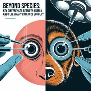
Cataract surgery is one of the most refined and standardized procedures in human ophthalmology. However, when we cross the species barrier and enter the world of veterinary ophthalmology, we find that this “routine” operation demands a distinct set of skills, considerations, and adaptations.

For ophthalmologists and veterinarians alike, understanding the nuanced differences between human and animal cataract surgery is essential—not just for successful outcomes, but also for appreciating the complexity of interspecies surgical practice.
Here, we explore the main divergences in technique, anatomy, and surgical environment that separate the two worlds.
1. Preoperative Evaluation: Systemic Stability Comes First
In human patients, preoperative clearance typically involves cardiovascular risk stratification and managing systemic comorbidities. In veterinary patients, the primary concern is ensuring the animal can safely tolerate general anesthesia, a necessity due to the impossibility of cooperation under topical or local anesthesia.
Furthermore, veterinarians must screen for ocular and systemic infections, including conditions involving the eyelids, ocular surface, or systemic immune status. These factors can contraindicate surgery or increase postoperative complications.
2. Ocular Anatomy: A Different Landscape
The structural anatomy of the animal eye differs substantially from the human eye, and this has direct surgical implications:
- Deeper anterior chamber: Common in canine patients, altering the ergonomics of intraocular manipulation.
- Larger lens volume: More mass to emulsify, requiring adjustments in phaco technique and settings.
- Minimal conjunctival exposure: Limits options for incisions, making the corneal incision the preferred one.
- Presence of the third eyelid (nictitating membrane): A unique challenge that can interfere with blepharostat positioning and surgical exposure.
These anatomical differences require both preoperative planning and intraoperative adaptation.
3. Surgical Setup: Ergonomics Matter More Than Ever
Whereas human surgical suites are optimized for the upright patient, veterinary cataract surgery demands more customized ergonomics:
- Adjustable surgical tables must accommodate a recumbent, anesthetized animal, with room for the phaco machine and microscope arms.
- Surgeon seating and microscope positioning must be precisely tailored to maintain comfort and precision over longer, technically demanding cases.
- Lateralization of the patient’s head is often necessary to achieve proper visualization and access to the corneal plane.
4. Surgical Steps: Familiar Yet Unpredictable
Capsulorhexis, a familiar yet critical step, presents significant challenges in veterinary surgery. Unlike the thin, predictable capsule in humans, the canine anterior capsule is:
- Thicker
- More elastic
- Less predictable in its tearing behavior
These factors demand superior manual control, a refined technique, and often, a greater reliance on viscoelastics to stabilize the anterior chamber.
Trypan blue staining becomes indispensable, given the difficulty in visualizing the anterior capsule in many animal patients.
5. Phacoemulsification: Same Technology, Different Tactics
Although modern phacoemulsification machines are used in both human and veterinary contexts, parameter settings must be adapted for the unique intraocular dynamics of animal eyes.
For instance, a typical veterinary case might begin with:
- Sculpting at 50% ultrasound power, 100 mmHg vacuum, 20 cc/min flow
- Quadrant removal with increased vacuum (400 mmHg), flow (35 cc/min), and a 40% burst ultrasound mode
Throughout the procedure, intraocular pressure stability is critical—particularly in the face of unpredictable chamber dynamics caused by the thicker lens and deeper anatomy.
6. IOL Implantation: Precision in a Challenging Space
In both fields, the goal remains the same: restore visual function with a well-centered, in-the-bag intraocular lens (IOL).
In veterinary patients, foldable lenses such as the Cristalens Loki (France) or AJL Vet Liocan (Spain) are often selected for their biocompatibility and ease of insertion. Use of cohesive viscoelastics is preferred to ensure stability during IOL placement.
7. Postoperative Management: Inflammation Is a Greater Threat
Postoperative inflammation tends to be more intense and less predictable in veterinary patients. Hence, intracameral injections of steroids and t-PA (tissue plasminogen activator) are often used proactively to mitigate fibrin formation and anterior segment complications.
The surgeon must also consider the owner’s ability to administer postoperative medications, which directly affects outcomes—a challenge not present in human cases.
Not Just a different Eye
Veterinary cataract surgery is not simply human cataract surgery applied to a smaller (or differently shaped) eye. It is a field with unique challenges that require customized solutions. For ophthalmic surgeons crossing into the veterinary world—or veterinarians integrating advanced surgical techniques—the key lies in recognizing anatomical, procedural, and logistical differences while leveraging shared knowledge and technology.
Interspecies expertise is not only possible—it’s a growing necessity in a world where animal visual quality is increasingly valued. Whether you’re a human ophthalmologist or a veterinary specialist, there is much to learn from each other on the surgical frontier of sight.
🧠 What you’ll gain:
✔️ Step-by-step mastery of phaco techniques
✔️ Hands-on simulation and case-based learning
✔️ Guidance from expert surgeons with experience in both human and animal ophthalmology
✔️ Immediate, real-world applications to your practice
🎯 Don’t leave surgical excellence to chance—invest in your skills and in your patients’ vision.
👉 Enroll now and become the cataract surgeon your patients deserve.
If you have any kind of interest please fill the following form and we will provide you with the information you need.
